


非创伤性股骨头坏死:外侧柱存留与股骨头塌陷的关系研究
2013-01-28 文章来源:中日友好医院 李子荣 我要说
骨科在线版权所有,如需转载请注明来自本网站
非创伤性股骨头坏死:外侧柱存留与股骨头塌陷的关系研究
李子荣 孙伟 史振才 赵凤朝* 王佰亮 张启栋 郭万首
基金项目:卫生部属(管)医院临床学科重点项目(2010-2012年度,152)
作者单位:100029北京,中日友好医院骨科、骨坏死与关节保留重建中心(赵凤朝:现在徐州医学院附属医院骨科)
通信作者:李子荣 E-mail: lzr96@hotmail.com
【摘要】
目的 追踪早期不同MRI或CT影像的股骨头坏死的最终结局,探明股骨头外侧柱存留与股骨头塌陷的关系。
方法 将股骨头冠状位正中层面划分为三柱(内侧、中央、外侧)。按初次检查或术前检查的股骨头MRI或CT,选用T1WI的冠状位正中层面图像,依据坏死灶占据外侧柱情况,分为三型。Ⅰ型:坏死灶占据中央柱和内侧柱、外侧柱完全存留;Ⅱ型:部分外侧柱存留(至少有外侧皮质存留),其余均被坏死灶占据;Ⅲ型:坏死灶占据整个外侧柱,坏死带穿透骨髓及外侧皮质。87例127髋ARCO Ⅰ期股骨头坏死,Ⅰ型37髋,Ⅱ型47髋,Ⅲ型43髋。非手术治疗,追踪3~8年(平均6.2年),至少检查3次。42例72髋,ARCO Ⅱ期股骨头坏死,术前Ⅰ型10髋,Ⅱ型32髋,Ⅲ型30髋。手术治疗(经股骨头颈开窗减压病灶清除,打压植骨),随访5~9年(平均7.1年)。
结果 未手术组,塌陷的股骨头,Ⅰ型4髋(10.8%),Ⅱ型20髋(42.6%),Ⅲ型41髋(95.3%)。塌陷的时间间隔,Ⅰ型5~7年,Ⅱ型2~7年,Ⅲ型0.5~3年。手术组,塌陷的股骨头,Ⅰ型无塌陷,Ⅱ型7髋(21.8%),Ⅲ型18髋(60.0%),塌陷的时间间隔,Ⅱ型2~5年,Ⅲ型2~3年。
结论 股骨头坏死是否会进展到塌陷与坏死灶是否累及股骨头外侧柱及累及程度密切相关,外侧柱存留者,塌陷率低,股骨头球形维持时间长,外侧柱完全累及者,多数股骨头将在较短时间内进展到塌陷。
【关键词】股骨头坏死; 塌陷; 外侧柱; 磁共振成像
The study on the relationship between the preserving of lateral pillar and collapse of the femoral head in patients with osteonecrosis
Li Zirong, Sun Wei, Shi Zhengcai, Zhao Fengchao, Wang Bailiang, Zhang Qidong, Guo Wanshou
Centre for Osteonecrosis and Joint-preserving & Reconstruction, China-Japan Friendship Hospital, Beijing, 100029, China
[Abstract]
Objective To inquire the final outcome of early stage of the osteonecrosis of the femoral head (ONFH) with different pictures of MRI or CT, to verify the relationship between the preserving of the lateral pillar and collapse of the femoral head.
Methods The mid-coronal section of the femoral head was divided into three pillars (medial, central and lateral). The mid-coronal section of the femoral head on MRI (T1WI) was selected. According to the site of the lateral pillar occupied by necrotic focus, the necrosis was divided into three types: type Ⅰ: the necrotic focus occupied the central and medial pillars, the lateral pillar preserved; type Ⅱ: the necrotic focus occupied the partial lateral pillar, the lateral pillar preserved partially (at least the cortical bone); type Ⅲ: the lateral pillar occupied by necrotic focus totally. Eighty-seven patients (127 hips) with ARCO stage Ⅰ ONFH were followed-up for 3 to 8 years (an average 6.2 years). All patients were nonoperative treatment. Forty-two patients (72 hips) with ARCO stage Ⅱ ONFH were followed-up for 5 to 9 years can average 7.1 years. All patients were carried out by debridement and operation with impacting bone graft.
Results In the nonoperative group there were 37 hips with type Ⅰ, 4 hips progressed to collapse (10.8%). Twenty hips progressed to collapse (42.6%) in the 37 hips with type Ⅱ. Forty-one hips progressed to collapse (95.3%) in the 43 hips with type Ⅲ. The duration from primary check to collapse was 5 to 7 years (type Ⅰ), 2 to 7 years (type Ⅱ) and 0.5 to 3 years (type Ⅲ). In the operative group, no hip with typeⅠprogressed to collapse , eighteen hips progressed to collapse in the 30 hips with type Ⅱ, seven hips progressed to collapse in the 20 hips with type Ⅲ. The duration from the time of operation to collapse was 2 to 5 years (type Ⅱ) and 2 to 3 years (type Ⅲ) respectively.
Conclusion ONFH whether progressed to the collapse or not is determined by preserving of the lateral pillar or not. The lateral pillar preserved, the femoral head will maintain the sphere. The more preserve the lateral pillar, the less collapse occurred. The more preserve lateral pillar, the longer maintain sphere.
[Key words] Osteonecrosis of the femoral head; Collapse Lateral pillar; Magnetic resonance imaging
大量的临床资料显示,未经有效治疗,多数坏死股骨头将进展到股骨头塌陷。股骨头一旦塌陷,则难以避免进展至髋关节骨关节炎,从而不得不接受人工关节置换(1-4)。股骨头塌陷是导致最终髋关节功能障碍的最主要原因。因此,预测坏死股骨头是否会进展到塌陷?经历多长时间会塌陷?能否在最大程度上阻止塌陷的发生均是研究的热点和重点。国外对股骨头坏死塌陷的预测已有许多方法,各有其优点和局限性,仍有改进的空间(5-9)。
选用ARCO分期坏死面积在中度及以上(10),应用皮质类固醇(corticosteroid,以下简称激素)引发股骨头坏死(osteonecrosis of the femoral head, ONFH)的严重急性呼吸综合征(severe acute respiratory syndrome, SARS)患者长期随访研究,以及对我科骨坏死中心收治的保存患者自身关节手术(joint-preserving operation,以下简称保头手术)中资料完整且获长期随访者的非创伤性ONFH(激素、酒精)的患者结果的研究,我们发现坏死股骨头是否会进展到塌陷与股骨头外侧柱存留与否及存留宽度密切相关。本文报告部分研究结果。
资料和方法
一、研究方法
1. 将股骨头标本按冠状位正中层面分为外侧柱、中央柱和内侧柱(图1),各柱宽度分别为30%、40%、30%(11)。
2. 依据SARS后ARCO分期Ⅰ期的ONFH患者(自然进展组)初次普查时的MRI或CT片,选用冠状位正中层面的T1WI(MRI)图像或冠状位二维重建的正中层面的(CT)图像,根据坏死灶占据外侧柱情况分为三型。Ⅰ型:坏死灶占据中央及内侧柱,外侧柱完全保留;Ⅱ型:部分外侧柱被坏死灶占据,部分保留(至少外侧皮质保留);Ⅲ型:坏死带穿透股骨头骨髓与外侧皮质,外侧柱无存留。(图2)
3. 保头手术患者均为ARCO分期的Ⅱ期(手术治疗组),依据术前MRI或CT,选用术前冠状位正中层面的T1WI(MRI)或冠状位重建正中层面的(CT)图像,按研究方法2逐例分型。
4. 对SARS患者随诊观察各型的塌陷率及塌陷与初次检查的间隔,对手术患者随诊记录各型塌陷率及塌陷与手术的时间间隔。
二、临床资料
1. 自然进展组:共87例127髋,初次检查时年龄21~59岁(平均33±9岁),男26,女61;双髋45例,右髋17例,左髋20例。
这些患者均在初次检查时(2003.12~2004.2)行3个月的非手术治疗。治疗项目包括中西药物(抗凝、活血化瘀、减慢骨吸收、促进骨修复、保护软骨等)、保护性负重、理疗、体疗、磁场治疗等。均获得至少3年(3~8年)随访,每年随访一次的随访,所有患者至少有3次(3~6次)随访。按前述方法分型,计Ⅰ型37髋,Ⅱ型47髋,Ⅲ型43髋。
2. 手术治疗组:我院骨科骨坏死中心2004.1~2006.7行保头手术的非创伤性ONFH,共42例72髋资料完整,术后6月,1年及以后每年随访,均获最少5年随访(5~8年)。男19,女23,手术时年龄22~54岁(平均30.7±6岁)。
手术方法:经髋关节前路改良Watson-Jones入路,显露髋关节前侧,股骨头颈交界处开窗,坏死灶清除,自体髂骨与人工骨混合打压植骨术。术后用减慢骨吸收及加速骨修复的中西药治疗6月~1年。保护性负重3~6月。详细技术见本中心发表的论文(12)。
术前坏死分型:Ⅰ型10髋,Ⅱ型32髋,Ⅲ型30髋。
三、统计学方法
采用SPSS17.0统计软件进行数据统计分析,计量资料以均数±标准差表示,计数资料采取X2检验及Fisher精细统计。
P>0.05为差异有统计学意义。
结果
一、 自然进展组
外侧柱完全存留的坏死(Ⅰ型),股骨头塌陷率低,即使发生塌陷,间隔时间也较长;外侧柱部分存留的坏死(Ⅱ型),股骨头塌陷率明显升高(与Ⅰ型相比,P=0.014),塌陷的间隔时间明显延长;坏死带穿透骨髓及外侧皮质,即所谓全股骨头型坏死(Ⅲ型),塌陷率高(与Ⅱ型相比,P=0.001),且间隔时间明显缩短(图3),各型之间差别均有统计学意义(表1)。
表1 各型股骨头坏死自然进展的塌陷率及塌陷间隔
Tab.1. The rate and duration of the collapse with natural history of ONFH in different types.
|
分型(MRI或CT) |
坏死数(髋) |
塌陷(髋,%) |
塌陷间隔(年) |
计数资料 |
P值 |
|
Ⅰ |
37 |
4(10.8) |
5-7 |
10.22(Ⅰ-Ⅱ间) |
0.014 |
|
Ⅱ |
47 |
20(42.6) |
2-7 |
57.75(Ⅰ-Ⅲ间) |
0.001 |
|
Ⅲ |
43 |
41(95.3) |
0.5-3 |
28.6(Ⅱ-Ⅲ间) |
0.001 |
二、 手术治疗组
外侧柱完全保留患者(Ⅰ型),保头手术后无股骨头塌陷,而外侧柱部分保留患者(Ⅱ型),手术后2~5年内21.8%进展到塌陷(Ⅰ型与Ⅱ型对比P=0.003),而坏死带穿透股骨头骨髓及皮质,即外侧柱完全无存的患者(Ⅲ型),手术后2~3年内有60%进展到股骨头塌陷(Ⅰ型与Ⅲ型对比P=0.008,Ⅱ型与Ⅲ型对比,P=0.008),各型之间塌陷率差别均有统计学意义。其进展规律与自然进展相同,但相同类型坏死的塌陷率明显降低,降低的比例也不一致,Ⅰ、Ⅱ型降低明显,Ⅲ型降低少,显示保头手术治疗对各型坏死均有不同疗效(表2)。
表2 各型股骨头坏死保头手术的塌陷率及塌陷间隔
Tab.2. The rate and duration of the collapse with joint-preserving operation of ONFH in different types.
|
坏死类型 |
坏死数(髋) |
塌陷数(髋,%) |
塌陷间隔(年) |
计数资料 |
P值 |
|
Ⅰ |
10 |
|
|
16.10(Ⅰ-Ⅱ间) |
0.003 |
|
Ⅱ |
32 |
7(21.8) |
2-5 |
—(Ⅰ-Ⅲ间) |
0.008 |
|
Ⅲ |
30 |
18(60.0) |
2-3 |
9.35(Ⅱ-Ⅲ间) |
0.002 |
注:计数资料为X2值,Fisher精确统计无计数值
讨论
临床观察非创伤性ONFH的患者的发展过程,发现不论是自然进展还是保头手术治疗的结局,最终总有部分股骨头发生塌陷,而部分股骨头仍可维持圆形。仔细研究对比股骨头坏死早期的MRI,发现有两种不同的图像。一种是坏死带仅限于股骨头骨髓,皮质骨或多或少存留(图2-1),而另一种为坏死带穿透股骨头骨髓与皮质(图2-2,3)。追踪这些患者,发现这两种不同MRI影像改变的坏死股骨头有明显不同的结局,前者股骨头长期保持圆形而不塌陷,CT扫描或X线摄片可显示股骨头皮质完整,坏死位于髓腔内;而后一种则股骨头塌陷变扁,CT扫描或X线摄片可显示软骨下骨折。两种不同MRI影像改变的股骨头坏死进展为不同结局引起了我们的思索。
Herring等(12)对大量儿童Legg-Perthes病患者进行了长期随访,发现坏死的股骨头骨骺外侧柱存留的高度与发育成熟后修复的股骨头圆形程度成正比,并据此提出了以外侧柱改变为基础的Legg-Perthes的Herring分型(12)。受此启发,联系到我们临床观察的发现,促使我们进一步研究股骨头坏死的塌陷与外侧柱存留及存留宽度的关系。本研究的SARS股骨头坏死患者的自然进展及保头手术治疗股骨头坏死患者的结局,显示股骨头坏死是否会进展到塌陷与外侧柱存留与否及存留宽度密切相关。与自然进展相比,手术治疗的结果显示,各型坏死的塌陷率均低于自然进展组,但手术不能完全避免股骨头塌陷。
早期骨坏死MRI影像的上述改变有其病理基础(13)。国内外多数学者均同意,非创伤性骨坏死(激素、酒精等)首先使骨髓内血管内皮细胞受损,继而引发骨髓内微血管栓塞。栓塞首先引发骨髓内成分(间充质细胞及脂肪细胞等)坏死,骨细胞紧接因缺氧而死亡,骨小梁变细而断裂(14-15)。不同坏死患者,血管栓塞可发生在股骨头供血网中的不同分支水平。主干栓塞,坏死体积大,分支栓塞,坏死体积小。股骨头的主要营养血管为上干骺动脉,此血管进入股骨头后,发生分支分别供应皮质、软骨下骨及骨髓。因此不同分支水平的血管栓塞引起不同类型的坏死,可在MRI上显示为不同影像,为我们判断坏死股骨头的外侧柱是否存留,以及存留宽度提供了可直视的图像。依此图像进行分型,预测股骨头是否会进展到塌陷,准确度高。
在股骨头坏死塌陷与外侧柱存留及存留宽度的关系深入研究的基础上,我们提出了以股骨头三柱结构为基础的股骨头坏死分型(中日友好医院分型(16)),此分型以三柱结构为解剖基础,根据MRI(ARCOⅠ~Ⅱ期)或CT(ARCOⅡ~Ⅲ期)冠状位正中层面影像改变(坏死灶占据三柱情况),分为A、B、C三大型,C型又分为C1、C2、C3三型,分别代表外侧柱存留情况(图4)。用此型对大量股骨头坏死患者进行自然进展或保头手术效果的研究,发现此分型应用的方便性,准确性均优于日本骨坏死调查班(JIC)提出的股骨头坏死分型(A、B、C1、C2四型)(5)。但仅限于本中心病例,期望同行用此分型法分析各自的病例,以获多中心研究的数据。
本研究的局限性为尚未进行外侧柱存留宽度与股骨头塌陷率与塌陷间隔时间之间的定量研究(17)。尚未就年龄、性别等因素是否影响结果进行研究。缺乏生物力学研究作理论支撑。
尽管如此,基于大量长期临床病例资料观察的结果或许更能反映骨坏死进展规律,用此分型判断股骨头坏死的预后,实施个体化治疗方案,或许可进一步提高治疗的针对性。
参考文献:
[1] Mont MA,Zywiel MG ,Marker DR et al. The Natural History of Untreated Asymptomatic Osteonecrosis of the Femoral Head: A Systematic Literature Review. J Bone Joint Surg (Am). 2010, 92(12): 2165-2170.
[2] Mont MA, Jones IC, Herngerfood DS. Nontraumatic osteonecrosis of the femoral head:ten years later. J Bone Joint Surg (Am). 2006, 88: 1117-1132.
[3] Lieberman JR, Berry DJ, Mont MA, et al. Osteonecrosis of the hip: management in the twenty-first century. J Bone Joint Surg (Am). 2002, 84: 834-853.
[4] Lieberman JR, Engstrom SM, Meneghini RM, et al. Which Factors Influence Preservation of the Osteonecrotic Femoral Head? Clin Orthop Relat Res. 2012, 470(2): 525-534.
[5] Sugano N, Kubo T, Takaoka K, et al. Diagnostic criteria for non-traumatic osteonecrosis of the femoral head: a multicentre study. J Bone Joint Surg Br. 1999, 81(4): 590-595.
[6] Koo KH, Kim R. Quantifying the extent of osteonecrosis of the femoral head: a new method using MRI. J Bone Joint Surg (Br). 1995, 77: 875-880.
[7] 李子荣,张念非,史振才,等.股骨头坏死塌陷的预测与治疗选择. 中华骨科杂志,2003, 23(4): 193-196.
[8] 史振才,李子荣,孙伟,等.计算机处理MR图像股骨头坏死体积测定与初步力学测试.中华放射学杂志. 2006, 40: 288-292.
[9] 赵凤朝,李子荣,张念非,等.坏死面积比例在预测股骨头塌陷中的价值.中华骨科杂志. 2005, 25: 520-523.
[10] Gardeniers JWM. A new international classification of osteonecrosis of the ARCO committee on terminology and classification. ARCO News Letter. 1992, 4: 41-46.
[11] Herring JA, Kim HT, Browne R. Legg-Calve-Perthes disease. Part I: Classification of radiographs with use of the modified lateral pillar and Stulberg classifications. J Bone Joint Surg (Am),2004,86(10):2103-2120
[12] Wang BL, Sun W, Shi ZC, et al. Treatment of nontraumatic osteonecrosis of the femoral head using bone impaction grafting through a femoral neck window. Int Orthop. 2010,34:635~639.
[13] 李子荣.股骨头坏死治疗方法选择的病理学基础.中华医学杂志,2007, 87(29):2021-2022
[14] Johnson EO, Soultanis KS, Panayotis N. Vascular anatomy and microcirculation of skeletal zones vulnerable to osteonecrosis: vascularization of the femoral head. Orthop Clin North Am. 2004, 35(3): 285-291.
[15] Drescher W, Li Haisheng, Lundgaard A, et al. Endothelin-1-induced femral head epiphyseal artery constriction is enhanced by long-term corticosteroid treatment. J Bone Joint Surg (Am),2006, 88: 173-179
[16] 李子荣,刘朝晖,孙伟,等. 基于三柱结构的股骨头坏死分型--中日友好医院分型. 中华骨科杂志,2012,32(6):515-520
[17] Ito H, Tanino H, Yamanaka Y, et al. Long-term results of conventional varus half-wedge proximal femoral osteotomy for the treatment of osteonecrosis of the femoral head. J Bone Joint Surg Br. 2012, 94(3):308-314.
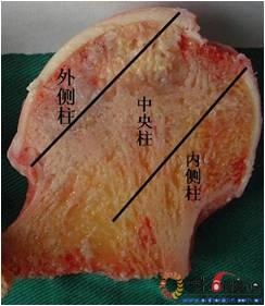
图1 股骨头冠状面三柱结构:外侧柱占30%,中央柱占40%,内侧柱占30%
Fig.1 Photograph of coronal section of the femoral head showing three pillars of the femoral head: lateral (30%), central (40%), medial (30%)
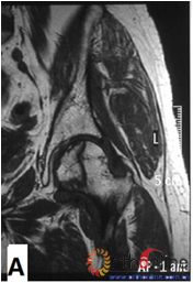

图2-1
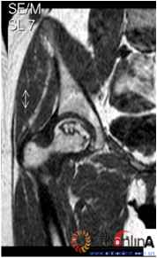

图2-2
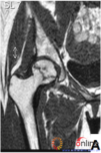
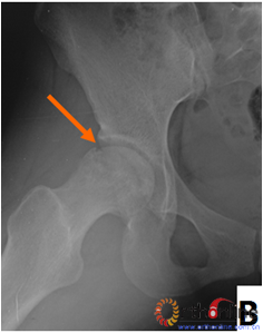
图2-3
图2 依据股骨头外侧柱存留情况,股骨头坏死的分型。(图2-1)分为Ⅰ型(外侧柱全部存留)(A)MRI图像显示;(B)7年后CT显示股骨头仍未塌陷;(图2-2)Ⅱ型(外侧柱部分保留)(A)MRI显示外侧皮质存留;(B)4年后CT显示股骨头仍未塌陷;(图2-3)Ⅲ型(坏死带穿透股骨头);(A)MRI显示坏死带;(B)2年后股骨头塌陷
Fig 2 According to preservation of the lateral pillar, the necrosis was divided into three types. (Fig 2-1)Type Ⅰ: Whole lateral pillar preserved (A) MRI showing; (B) no collapse occurred seven years later by CT showing. (Fig 2-2)Type Ⅱ: Preservation of the partial lateral pillar, (A) MRI showed the preservation of the lateral cortical bone; (B) no collapse of the femoral head four years by CT. (Fig 2-3)Type Ⅲ: the necrotic line pass through the cortical bone and bone morrow ;(A) MRI showed the necrotic line; (B) collapse of the femoral head occurred two year later.


图3 各型坏死打压植骨的结果,(A)Ⅰ型(右侧)和Ⅱ型(左侧)术前MRI显示外侧柱存留;(B)术后7年随访,股骨头维持外形,关节功能好;(C)Ⅲ型(双侧)术前MRI显示外侧柱破坏;(D)术后5年,右侧股骨头维持外形,左侧塌陷
Fig 3 The results of different osteonecrotic type by impacting bone graft,(A)Type Ⅰ(right),type Ⅱ(left) MRI showed the preservation of the lateral pillar preoperatively; (B) Seven years after operation, the femoral head still maintain sphere, hip function is excellent; (C) Type Ⅲ (both side) MRI showed the involved of lateral pillar by necrosis; (D)Five years after operation, the femoral head still maintain sphere in right side, collapse occurred in left side.
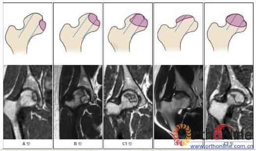
图4 依据三柱理论的CJFH 股骨头坏死分型示意图及MRI 影像。A 型:坏死灶占据内侧柱,B 型:坏死灶占据中央柱和内侧柱,C1 型:坏死灶占据三柱,但外侧柱有部分存留,C2 型:坏死灶占据外侧柱及部分中央柱,C3 型:坏死带穿透整个股骨头三柱皮质及骨髓
Fig.4 Schematic diagram and MRI of China-Japan Friendship Hospital (CJFH) classification for osteonecrosis of the femoral head based on three pillars. Type A: the necrosis involved the medial pillar. Type B: the necrosis involved both medial and central pillars. Type C1: the necrosis involved the three pillars but the partial lateral pillar preserved. Type C2: the necrosis occupied all lateral pillar and partial central pillar. Type C3: the necrosis involved the three pillars including the cortical bone and marrow.
 京公网安备11010502051256号